Research
Hysteroscopy vs. Ultrasound: Endometrial Examination Methods
Explore the differences between hysteroscopy and ultrasound for endometrial health, especially in women with PCOS, and understand when to use each method.
August 8, 2025
·
12
Explore the differences between hysteroscopy and ultrasound for endometrial health, especially in women with PCOS, and understand when to use each method.
Hysteroscopy and ultrasound are two methods used to examine the uterus, especially for women with PCOS who face higher risks of endometrial issues like hyperplasia or cancer. Here's a quick breakdown:
| Factor | Hysteroscopy | Ultrasound |
|---|---|---|
| Visualization | Direct view of the uterus | Images via sound waves |
| Biopsy Capability | Yes | No |
| Procedure Time | 5–30 minutes | 15–30 minutes |
| Anesthesia | May be required | None |
| Recovery Time | 1–2 days | Immediate |
| Risks | Rare (e.g., infection, bleeding) | Minimal |
Ultrasound is ideal for initial screening, while hysteroscopy is better for detailed diagnosis and treatment. Combining both methods ensures thorough care, especially for PCOS patients.
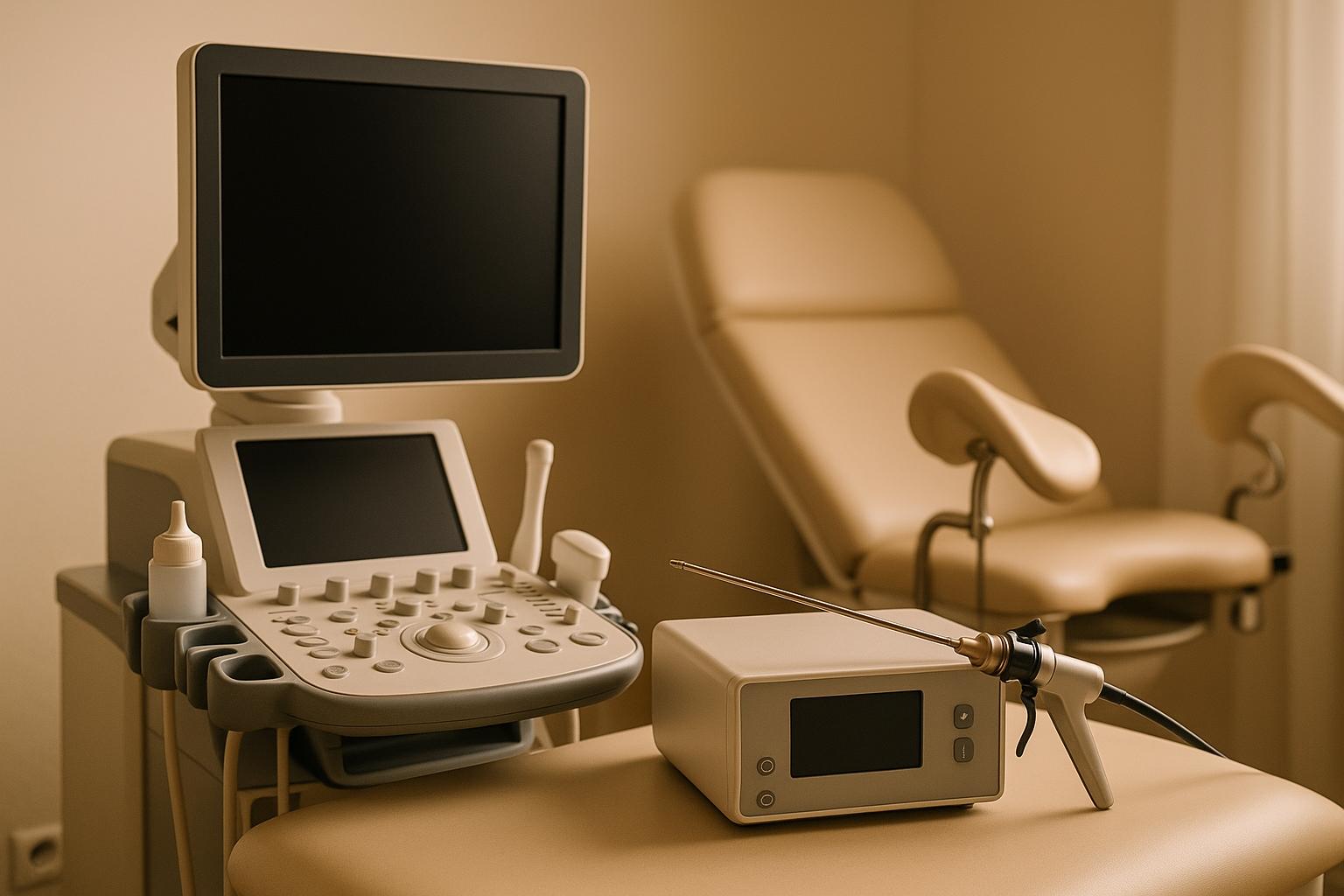
Hysteroscopy is often regarded as the gold standard for diagnosing and treating intrauterine issues. By providing a direct view of the uterine cavity, it plays a key role in monitoring the endometrium, particularly in women with PCOS.
Hysteroscopy involves inserting a slim, lighted instrument called a hysteroscope through the vagina and cervix into the uterus - making it a procedure that requires no incisions.
"Hysteroscopy involves inserting a rigid or flexible hysteroscope through the cervical canal into the uterus and then using distending media to allow for complete visualization of the endometrial cavity, allowing for minimally invasive diagnosis and surgical management of endocervical and intrauterine pathology." - Jessica F. Moore, Author
To ensure a clear view, the uterine cavity is gently expanded using fluid or gas, allowing the doctor to examine the endometrial lining in detail. This step is critical for detecting even small abnormalities that might not be visible through other diagnostic methods. These procedural elements not only enhance visibility but also highlight the clinical value of hysteroscopy.
The procedure can serve two purposes: diagnosis (to identify issues) or treatment (to address problems during the same visit). For the best results, it’s usually performed during the first week after menstruation. Depending on the complexity, the process can take anywhere from 5 minutes to over an hour.
Hysteroscopy is versatile, performed in various clinical settings with anesthesia options tailored to ensure patient comfort.
Hysteroscopy offers a range of advantages, particularly for women with PCOS. One of its most important benefits is its ability to detect mild intrauterine abnormalities that might go unnoticed on an ultrasound. Research shows that 20-40% of women with normal transvaginal ultrasound results actually have mild issues revealed during hysteroscopy. This is especially critical for women with PCOS, who face a heightened risk of endometrial problems.
If abnormalities are found, hysteroscopy allows for targeted biopsies or the complete removal of problematic tissue during the same session. This dual capability makes it possible to diagnose and treat conditions like endometrial polyps and other structural issues in a single appointment.
For women trying to conceive, hysteroscopy can boost live birth rates, particularly when undergoing IVF. Additionally, in-office hysteroscopy provides the added benefits of convenience, quicker recovery, and higher patient satisfaction.
While hysteroscopy is highly effective, it does come with some limitations. Since it involves inserting instruments into the uterus, it is more invasive than an ultrasound. Patients may experience pain or discomfort, which can vary based on the procedure’s duration and individual pain tolerance.
Certain conditions, such as pregnancy or active pelvic infections, make hysteroscopy unsuitable. Your doctor will screen for these before scheduling the procedure.
Although complications are rare, there are potential risks, including infection, bleeding, or injury to the uterus or nearby organs. About 1 in 200 women undergoing hysteroscopy with local anesthesia may feel faint during the procedure. The risk of uterine perforation is estimated to be between 0.002% and 1.7%, according to the Royal College of Obstetricians and Gynaecologists.
After the procedure, contact your doctor if you experience fever, severe abdominal pain, or heavy bleeding. Most women recover quickly, but discussing pain management options with your healthcare provider beforehand is always a good idea.
Ultrasound is a non-invasive and essential diagnostic tool for evaluating the endometrium, especially in women with PCOS. This imaging method is pivotal for monitoring endometrial thickness and detecting abnormalities, given the heightened risk of endometrial hyperplasia in this population.
The primary technique for endometrial evaluation is transvaginal ultrasound (TV-US), which offers a clearer and more detailed view compared to transabdominal methods. During this procedure, a specialized probe is inserted vaginally to examine the uterus and ovaries.
One key aspect of this exam is measuring endometrial thickness. Research shows that an endometrial thickness under 7 mm effectively rules out hyperplasia in women with PCOS. This is particularly relevant, as studies reveal that 35.7% of women with PCOS experiencing infertility due to anovulation are diagnosed with endometrial hyperplasia.
For women with regular cycles, the test is best performed during the early follicular phase. For those with irregular cycles, it can be done randomly or 3–5 days after progesterone-induced bleeding.
"Although TVU represents a practical approach for the initial evaluation of uterine pathologies, a hysteroscopy examination would be necessary in most of the suspicious cases. Hysteroscopy seems to offer better diagnostic value for uterine pathologies in general, and uterine polyps in particular."
– Ali Babacan, Department of Obstetrics and Gynecology, GATA Haydarpasa Training Hospital, Istanbul, Turkey
This highlights the importance of ultrasound as a first step in assessing endometrial health, though additional procedures like hysteroscopy may be needed for more detailed investigations.
Ultrasound stands out as a practical choice for endometrial assessment due to its non-invasive nature. The procedure is generally painless, doesn’t require anesthesia, and allows patients to resume normal activities immediately. Its widespread availability and affordability make it a convenient option for early screening.
Transvaginal ultrasound is particularly reliable for initial evaluations, with a 99% negative predictive value for detecting early endometrial cancer. Additionally, when the endometrial thickness is less than 4.0 mm, the likelihood of endometrial cancer drops to less than 1%.
Beyond the endometrium, ultrasound also plays a role in assessing ovarian characteristics linked to PCOS. It can identify polycystic ovaries - present in over 90% of PCOS cases - and evaluate key factors like follicle count and ovarian volume.
While ultrasound offers many advantages, it does have its limitations. Unlike hysteroscopy, it doesn’t provide direct visualization of the uterine cavity, which may lead to subtle abnormalities being overlooked. Additionally, ultrasound cannot perform biopsies, so further testing might be required for a definitive diagnosis.
Its sensitivity for detecting certain ovarian features can also be inconsistent. For example, about 30% of patients with hormonal evidence of polycystic ovaries may have normal ovarian size on ultrasound, and fewer than half of those with increased ovarian volume display the classic pattern of multiple small peripheral follicles.
The accuracy of ultrasound heavily depends on the skill of the operator. In some cases, such as with women who have obesity, image quality can be compromised. Advanced techniques like 3D ultrasound may be necessary to achieve better visualization in these situations.
This section dives into the practical differences between hysteroscopy and ultrasound, particularly in managing PCOS. Understanding these distinctions can help in making informed choices for monitoring the endometrium in PCOS patients.
Hysteroscopy allows for direct visualization of the uterus, making it easier to spot subtle changes that ultrasound might overlook. Meanwhile, 3D ultrasound serves as an excellent initial screening tool, boasting up to 90% diagnostic accuracy.
When comparing the two methods, diagnostic accuracy varies. For example, transvaginal sonography (TVS) shows an 84.5% sensitivity and 98.7% specificity in detecting uterine cavity abnormalities, along with a 98.0% positive predictive value and an 89.2% negative predictive value. However, studies reveal that 20–40% of individuals with normal TVS results had mild intrauterine conditions detectable only through hysteroscopy.
Hysteroscopy stands out because it not only detects abnormalities but also allows for biopsy sampling, which is crucial for identifying endometrial disorders in PCOS patients.
Here’s a quick comparison of key factors:
| Comparison Factor | Hysteroscopy | Ultrasound |
|---|---|---|
| Visualization | Direct view of the uterine cavity | Indirect imaging via sound waves |
| Biopsy Capability | Enables targeted biopsies | Not possible |
| Procedure Time | 5–30 minutes | 15–30 minutes |
| Anesthesia Required | May require local, regional, or general | None required |
| Recovery Time | 1–2 days for normal activities | Immediate return to activities |
| Complication Rate | Less than 1% | Minimal to no known risks |
When it comes to detecting abnormalities, hysteroscopy excels at spotting small lesions (1–2 mm) and is highly effective for identifying endometrial polyps, submucous myomas, and intrauterine adhesions. On the other hand, 3D ultrasound is particularly reliable for detecting improperly positioned intrauterine devices, with a sensitivity of 93.3% and an accuracy of 86.2% compared to hysteroscopy.
These differences highlight when each method is most appropriate.
Ultrasound serves as an excellent first-line, non-invasive screening tool, especially for measuring endometrial thickness. Hysteroscopy, however, is better suited for cases where ultrasound findings are abnormal or when direct visualization and biopsy are required.
Even in cases where ultrasound results appear normal, hysteroscopy may still be beneficial. For example, one study found that 17.1% of PCOS infertile women with normal endometrial thickness on ultrasound had disordered proliferative endometrium.
For women with PCOS in their late reproductive years, hysteroscopy combined with an endometrial biopsy is particularly important. This is especially true for those experiencing prolonged amenorrhea, regardless of ultrasound results. The increased risk of premalignant and malignant endometrial polyps in premenopausal women with PCOS further underscores the importance of this approach.
Advancements in 3D ultrasound technology have also shaped clinical practices. With its high diagnostic accuracy, many specialists now recommend 3D ultrasound as the initial investigation for suspected uterine abnormalities, reserving hysteroscopy for confirmation and treatment.
While ultrasound is more cost-effective and non-invasive, hysteroscopy offers the advantage of providing a definitive diagnosis and treatment, making it a valuable tool despite its higher cost and minor discomfort.
Deciding between hysteroscopy and ultrasound for assessing endometrial health in PCOS requires a tailored approach based on clinical needs and available resources.
Start with a thorough clinical evaluation. Studies suggest that an endometrial thickness greater than 8.5 mm, or being older than 25.5 years, indicates a higher risk of endometrial disease. For those with irregular cycles, the risk increases with an endometrial thickness over 7 mm or intermenstrual intervals exceeding three months.
Risk assessment is critical, as PCOS significantly raises the likelihood of endometrial hyperplasia (21.4%) and cancer (1.7%). Chronic anovulation in PCOS results in prolonged exposure to unopposed estrogen, further heightening these risks.
For PCOS patients experiencing abnormal bleeding, hysteroscopy is recommended to check for polyps, hyperplasia, or cancer. However, ultrasound remains the first-line screening tool due to its non-invasive nature and affordability.
In cases of infertility, hysteroscopy with biopsy should be prioritized, even if ultrasound results appear normal. Research underscores the limitations of hysteroscopy alone in detecting certain endometrial issues:
"Hysteroscopic evaluation alone of infertile women without histological studies might not be able to detect probable endometrial pathologies such as micropolyps." - Amooee et al.
Patient preferences also play a role. Ultrasound is quick, non-invasive, and doesn’t require sedation, while hysteroscopy involves sedation and a short recovery period. Addressing these factors can improve patient comfort and acceptance.
By combining clinical assessments with patient-centered care, healthcare providers can ensure the most effective and personalized approach to endometrial health in PCOS, reducing long-term risks.

For additional guidance, PCOSHelp offers a wealth of resources designed to support women in managing their endometrial health. This platform provides detailed guides on fertility strategies, a vital resource for women incorporating endometrial evaluations into their fertility plans.
PCOSHelp’s tools for symptom tracking help users monitor menstrual irregularities and recognize when professional evaluation might be necessary. These resources empower women to communicate more effectively with their healthcare providers about potential concerns.
The site also emphasizes lifestyle adjustments for preventive care. From dietary tips to stress management techniques, PCOSHelp focuses on promoting hormonal balance, which is crucial for maintaining healthy endometrial function.
For those managing medications like Metformin or Spironolactone, PCOSHelp provides practical advice on how these treatments can influence endometrial health. Understanding these interactions helps women make well-informed decisions about their care.
Beyond immediate medical concerns, PCOSHelp highlights self-care strategies for long-term health. This aligns with clinical recommendations that stress the importance of comprehensive PCOS management in preventing complications related to endometrial health.
Hysteroscopy and ultrasound play pivotal roles in monitoring endometrial health in women with PCOS, working together rather than in isolation. Their combined use enhances clinical decision-making and ensures more accurate management of this condition.
Ultrasound serves as a non-invasive starting point, efficiently screening and measuring endometrial thickness. However, it’s worth noting that 20–40% of normal ultrasound findings may still conceal mild intrauterine abnormalities. This is where hysteroscopy proves indispensable. By providing direct visualization of the uterine cavity and enabling precise biopsies, hysteroscopy can uncover issues that ultrasound might overlook. For example, studies involving women with PCOS and seemingly normal ultrasound results revealed that hysteroscopic biopsies identified various conditions, including proliferative endometrium (71.67%), disordered proliferative endometrium (18.33%), chronic endometritis (3.33%), and secretory endometrium (6.67%).
In clinical practice, a step-by-step approach is often employed. Ultrasound is typically the first tool used for screening, followed by hysteroscopy when specific concerns arise - such as infertility, abnormal bleeding, or recurrent pregnancy loss. This sequential method ensures diagnostic accuracy while avoiding unnecessary invasive procedures.
For women with PCOS, this combined strategy is particularly critical due to their higher risk of endometrial complications. Hysteroscopy’s ability to detect subtle abnormalities, like micropolyps or chronic endometritis, can be key to guiding appropriate treatments and addressing reproductive challenges.
Rather than viewing ultrasound and hysteroscopy as competing options, they should be seen as complementary tools. Ultrasound provides a broad overview, acting as the initial guide, while hysteroscopy offers the detailed insights needed for conclusive diagnoses and effective treatment planning. Together, they form a powerful duo in the management of PCOS-related endometrial health.
Hysteroscopy is frequently chosen over ultrasound for examining the endometrium in women with PCOS because it offers direct visualization of the uterine cavity. This means it can more accurately identify abnormalities like polyps, hyperplasia, or lesions that might not be detected through ultrasound.
Another advantage of hysteroscopy is that it allows for targeted biopsies during the procedure. This enhances diagnostic precision, especially when identifying subtle endometrial issues that could impact fertility or uterine health. As a result, it serves as an essential tool for a thorough evaluation.
Recovery after a hysteroscopy is generally straightforward but may come with some mild discomfort. It's common to experience light cramping and spotting for up to a week. While most people can get back to their regular routines within 24 hours, certain precautions - like avoiding strenuous activities or using tampons - might be recommended for a few days. The cramping often feels similar to menstrual pain and can usually be eased with over-the-counter pain relievers. Always stick to the recovery guidelines provided by your healthcare provider.
On the other hand, ultrasound procedures typically involve little to no recovery time. Most individuals feel completely fine within a day or two, if not immediately. Side effects are rare and usually limited to minor issues like temporary redness or slight discomfort. Patients can usually return to their normal activities right away, making ultrasound a less invasive and quicker option for examining the endometrium.
For those managing PCOS, a hysteroscopy might be suggested instead of an ultrasound when there's a need to investigate intrauterine issues like a thickened endometrium, polyps, or unusual lesions. Unlike an ultrasound, hysteroscopy provides a direct view of the uterine lining and allows for a biopsy if necessary, offering clearer and more precise information.
This approach is especially helpful when ultrasound findings are inconclusive or if symptoms such as ongoing abnormal uterine bleeding persist. Since PCOS can heighten the risk of conditions like endometrial hyperplasia or even cancer due to prolonged exposure to estrogen, hysteroscopy becomes an important tool for diagnosis in these situations.