Research
Diagnostic Tools for Endometrial Polyps
Explore the key diagnostic tools for endometrial polyps, their accuracy, invasiveness, and how they impact treatment decisions.
July 16, 2025
·
11
Explore the key diagnostic tools for endometrial polyps, their accuracy, invasiveness, and how they impact treatment decisions.
Endometrial polyps are common, affecting about 25% of women, though most cases are symptom-free. These growths can cause abnormal uterine bleeding (20–40% of cases) and contribute to infertility (35% of cases). While the risk of cancer is low (0.5–5.4%), it increases for women with conditions like PCOS, who face a 2.75x higher chance of developing malignant polyps. Early and accurate diagnosis is essential for addressing symptoms, preserving fertility, and mitigating risks.
Here’s a quick overview of the main diagnostic tools:
Each method has strengths and limitations, and combining tools often yields the best results. Your healthcare provider can guide you toward the most appropriate diagnostic approach based on your symptoms and risk factors.
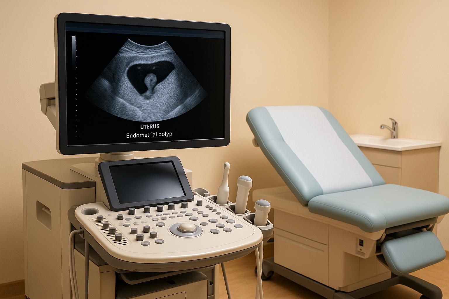
Transvaginal ultrasound is often the first choice for detecting endometrial polyps. It’s widely used because it’s accessible, non-invasive, and relatively affordable. This technique involves inserting a transducer into the vagina to create detailed images of the uterine cavity, making it an essential tool when symptoms like abnormal bleeding suggest polyps might be present.
The reliability of TVUS largely hinges on the skill of the examiner. Studies show significant variation in its accuracy, with sensitivity ranging from 39.8% to 89.6% and specificity from 39.1% to 72.7% when compared to hysteroscopy or histological findings. When performed by less experienced practitioners, TVUS tends to yield lower accuracy, which is why hysteroscopy is often recommended in cases where results are unclear.
This variability highlights the importance of pairing TVUS with other diagnostic methods for a more thorough evaluation.
TVUS is particularly effective at identifying endometrial polyps, which appear as hyperechogenic lesions with smooth edges within the uterine cavity. These may look like a focal mass or a general thickening of the endometrium. Adding color-flow Doppler technology can enhance detection by identifying the single feeding vessel characteristic of polyps, with Power Doppler improving sensitivity to nearly 97%.
The best time for a TVUS is around the 10th day of the menstrual cycle when the endometrium is at its thinnest. This timing ensures clearer imaging of any abnormal growths.
Such precision allows for more targeted follow-up evaluations.
TVUS is generally well-tolerated and causes less discomfort than a standard pelvic exam. The transducer used is smaller than a typical speculum, making the procedure minimally invasive.
"While no one looks forward to undergoing a transvaginal ultrasound exam, the procedure generally does not cause any physical discomfort, and it tends to be less invasive than a typical annual pelvic exam."
Patients are encouraged to share any discomfort during the procedure and may request a female technologist if that makes them more comfortable. Despite its limitations, TVUS remains a crucial diagnostic tool and a key step in evaluating endometrial polyps.
Sonohysterography (SIS) enhances transvaginal ultrasound by using saline to expand the uterus, offering a clearer view of endometrial polyps.
When it comes to identifying endometrial polyps, sonohysterography outperforms standard transvaginal ultrasound. According to meta-analyses and systematic reviews, the sensitivity of sonohysterography ranges from 86% to 87%, with specificity between 81% and 86% - results that are nearly on par with hysteroscopy . Beyond just numbers, this method provides a detailed look at uterine anatomy, offering valuable insights.
The saline infusion plays a key role in creating the ideal conditions for examining polyps in detail. This technique allows for precise identification of the number, size, location, and structure of polyps - whether they are pedunculated or sessile . Such detailed imaging is essential for planning treatment, as the characteristics of polyps often dictate the surgical approach.
Sonohysterography is a minimally invasive procedure that most patients find easy to tolerate. It involves inserting a slim ultrasound probe into the vagina and threading a thin catheter through the cervix into the uterus. Saline is then introduced to expand the uterine cavity, making the endometrial lining easier to examine . The entire process takes less than 30 minutes and is typically scheduled after menstruation but before ovulation . While some patients may experience mild cramping, it’s usually brief. Complications are rare, with infection occurring in just 0.95% of cases. Thanks to its high accuracy and safety, sonohysterography serves as an excellent middle ground between basic ultrasound and more invasive surgical options.
Hysteroscopy stands as the go-to method for diagnosing endometrial polyps, allowing a direct look into the uterine cavity using a slim, lighted telescope.
Compared to earlier imaging techniques, hysteroscopy delivers a higher level of accuracy. It boasts a sensitivity of 90% and a specificity of 93%, far surpassing standard ultrasound methods. When paired with a targeted biopsy under direct visualization, sensitivity increases to 35.3%–36.8%, compared to around 29.2% achieved with blind biopsy techniques. This precision is invaluable for assessing polyp characteristics more effectively.
Hysteroscopy provides a complete view of the endometrial cavity, making it possible to accurately determine the size, number, and type of polyp attachment. Polyps may either have a broad base (sessile) or be attached by a stalk (pedunculated). Since polyps are often located in the uterine fundus and appear as multiple growths in about 20% of cases, this comprehensive visualization is crucial for planning treatment.
While hysteroscopy is considered moderately invasive, it has a strong safety record, with complications occurring in fewer than 1% of cases. During the procedure, the uterine cavity is expanded using saline or carbon dioxide to improve visibility. Office-based hysteroscopy is a convenient option, performed on an outpatient basis without the need for general anesthesia, and typically causes only mild cramping. This approach not only ensures safety but also allows for therapeutic interventions during the same session.
One of hysteroscopy's key advantages is its ability to diagnose and treat at the same time. Polyps can be identified and removed during the procedure, with hysteroscopic polypectomy relieving symptoms in 75%–100% of cases. This method is both effective and cost-efficient, offering a streamlined solution for patients.
Diagnosing endometrial polyps effectively requires a combination of imaging and tissue sampling. While an endometrial biopsy provides valuable insights into tissue pathology, it does not offer a clear visual assessment of the uterine lining.
An endometrial biopsy involves extracting a small sample of the uterine lining using a thin tube inserted through the cervix. This procedure is typically recommended for women over 45 experiencing abnormal bleeding, younger women with a high risk of uterine cancer, or those with postmenopausal bleeding.
Endometrial biopsy is highly sensitive for detecting endometrial cancer, with a sensitivity rate of 96%. However, its ability to identify polyps is more limited. When performed blindly, the sensitivity for detecting polyps is about 29.2%, with a specificity of 60%. Using hysteroscopic guidance slightly improves sensitivity to around 35.3–36.8%, but specificity remains low at 33.3–50%. Larger, more diffuse, or multiple lesions are more likely to be detected than smaller, isolated polyps. Office-based biopsies generally have lower accuracy for identifying polyps compared to tissue obtained during surgical procedures. A meta-analysis also highlighted that in women with postmenopausal bleeding, the sensitivity of endometrial sampling for detecting issues like cancer, atypical hyperplasia, or polyps is lower than previously assumed. This suggests that further diagnostic steps may be necessary even after a benign biopsy result. These limitations emphasize the importance of combining biopsy results with imaging for a more thorough evaluation.
Unlike imaging techniques, an endometrial biopsy does not provide visual information about polyps. Since the procedure is performed without direct visualization, it cannot determine the size or location of polyps. Even when hysteroscopic guidance is used, the biopsy remains a sampling tool rather than a method for visual assessment. As a result, blind tissue sampling should only be considered when hysteroscopic treatment options are unavailable.
The procedure is relatively quick, taking about 10 minutes and typically performed in an outpatient setting without anesthesia. Some patients may experience mild cramping or light bleeding, with rare cases of infection or uterine perforation. To reduce discomfort, pre-procedure NSAIDs and topical anesthetics are often recommended. Patients are advised to avoid douching, using tampons, or engaging in sexual activity for 2–3 days following the procedure. However, it’s worth noting that around 31% of biopsies result in insufficient tissue samples for diagnosis, which can limit the effectiveness of the procedure.
When evaluating diagnostic tools, it's clear that each method has distinct advantages, drawbacks, and specific scenarios where it shines. Factors like accuracy, invasiveness, and treatment potential play a central role in determining the best approach for a given case.
The effectiveness of these methods varies significantly. Sonohysterography stands out with a perfect sensitivity and specificity of 100%. Transvaginal ultrasound (TVUS) follows closely, with a sensitivity of 88.3% and specificity of 91.2%. Hysteroscopy, while offering direct visualization, shows slightly lower sensitivity (83.9%) and specificity (63.0%) for detecting endometrial polyps. These numbers highlight the strengths and weaknesses of each tool, paving the way for more informed clinical decisions.
The ability to both visualize and treat polyps differs across methods:
The invasiveness of each method directly impacts patient recovery times:
Below is a comparison of key diagnostic parameters for each method:
| Method | Sensitivity for Polyps | Specificity for Polyps | Visualization Quality | Treatment Capability | Recovery Time |
|---|---|---|---|---|---|
| Transvaginal Ultrasound | 88.3% | 91.2% | Basic imaging | None | None |
| Sonohysterography | 100% | 100% | Excellent cavity outline | None | Immediate |
| Hysteroscopy | 83.9% | 63.0% | Direct visual assessment | Immediate treatment | ~24 hours |
| Endometrial Biopsy | 29.2% | N/A | No visualization | None | Minimal |
Choosing the right diagnostic method depends on the clinical presentation and the patient's unique circumstances. Transvaginal ultrasound is often the first step in screening for uterine abnormalities. If an issue is detected, sonohysterography can provide more detailed imaging to guide decisions about whether hysteroscopy is needed. This stepwise approach ensures a balance between thoroughness and efficiency.
Costs vary depending on the method. Transvaginal ultrasound is typically the most affordable and widely accessible option, making it an ideal first-line test. Hysteroscopy, on the other hand, is more costly due to the need for anesthesia and specialized facilities. However, its ability to combine diagnosis and treatment in one procedure can make it a more economical choice when intervention is required.
No diagnostic method is without flaws. For example, while endometrial biopsy is highly effective at detecting cancer (up to 96% sensitivity), it performs poorly for identifying polyps, with a sensitivity of just 29.2% when done blindly. Transvaginal ultrasound, though generally reliable, has reduced sensitivity for certain conditions, such as uterine adhesions or a unicornuate uterus, where sensitivity can drop as low as 35%. These limitations often make it necessary to combine multiple diagnostic tools to ensure a comprehensive evaluation. This multi-faceted approach helps clinicians craft tailored strategies for diagnosing and managing endometrial polyps effectively.
Diagnosing endometrial polyps requires a thoughtful approach that balances precision, patient comfort, and efficiency. The four main diagnostic tools - transvaginal ultrasound (TVUS), sonohysterography, hysteroscopy, and endometrial biopsy - each play a unique role in the evaluation process.
TVUS often serves as the first step, providing an initial overview. For a more detailed look, sonohysterography enhances visualization, helping to refine the diagnosis. When both direct visualization and immediate treatment are necessary, hysteroscopy becomes the go-to option, offering a clear view and the potential for simultaneous intervention. While endometrial biopsy may not be as effective at detecting polyps directly, it remains critical for assessing cancer risk in specific patients.
The choice of diagnostic method is not one-size-fits-all and should be tailored to the individual. For patients who are postmenopausal, over 60, experiencing postmenopausal bleeding, or taking tamoxifen, the risk of malignant polyps is higher, often warranting a more aggressive evaluation. On the other hand, asymptomatic patients with a low likelihood of malignancy might be managed with careful observation. Although cancerous transformation is uncommon, it’s essential to weigh patient-specific factors and symptoms to determine the urgency and type of diagnostic approach.
If you’re experiencing abnormal bleeding, fertility concerns, or postmenopausal symptoms, consulting your healthcare provider is key. They can help assess your risk factors and recommend the most appropriate diagnostic pathway, ensuring a thorough and personalized evaluation.
When it comes to diagnosing endometrial polyps, the choice of method hinges on factors like how precise and dependable the tool is. Transvaginal ultrasonography (TVUS) is usually the go-to option. Why? It’s non-invasive, easy to access, and does a solid job of spotting polyps.
If a clearer picture is needed or the initial results leave some uncertainty, hysteroscopy with guided biopsy is often the next step. This method allows doctors to directly examine the uterine cavity, making it the most trusted approach for a definitive diagnosis.
The decision can also be shaped by the patient’s symptoms, potential risk factors, and the need for a more detailed evaluation. These elements help guide the choice of the most suitable diagnostic tool.
Women with PCOS (Polycystic Ovary Syndrome) face a heightened risk of developing endometrial polyps, largely due to prolonged exposure to estrogen and factors like obesity. These hormonal imbalances can alter the uterine lining, making polyps or other endometrial issues more likely.
To identify endometrial polyps, doctors often rely on ultrasound or hysteroscopy, with hysteroscopy offering the most precise results. Early detection through these methods is crucial, as women with PCOS are more vulnerable to complications involving the endometrium.
Healthcare providers often rely on a combination of diagnostic methods to thoroughly evaluate endometrial polyps. Tools like transvaginal ultrasound and color Doppler imaging are used to identify abnormalities, while hysteroscopy with biopsy allows for a detailed examination and confirms the diagnosis.
By integrating these techniques, doctors can get a clearer picture, distinguish polyps from other conditions such as hyperplasia or cancer, and create an effective treatment plan tailored to the patient. This approach reduces uncertainty and supports more precise care.